Cookie information

Cookies are small files installed on your computer or smartphone. They allow us to store information about your navigation on our site.

Choose cookies Accept all cookies

Privacy Policy

Accept all cookies close

Cookie information

Olea Medical uses three types of cookies:

  • Session or preference cookies, which are essential for navigation and the good functioning of the site
  • Audience measurement cookies and other tracers used to establish site audience statistics

These cookies are configured according to the criteria for exemption from consent as defined by the CNIL

CookieTypeDescription
Service supplyStores consent for each "tracker" type cookie.
Service supplyStores authentication related to case report downloads
User authenticationCustomer area only. This cookie will not be set if you do not log in to your account.
Service supplyStores the current language
TrackingGoogle analytics

Neuroimaging APT CEST V.1.0 imaging solution
  • MRI

Mapping peptides and proteins to detect molecular changes in the brain

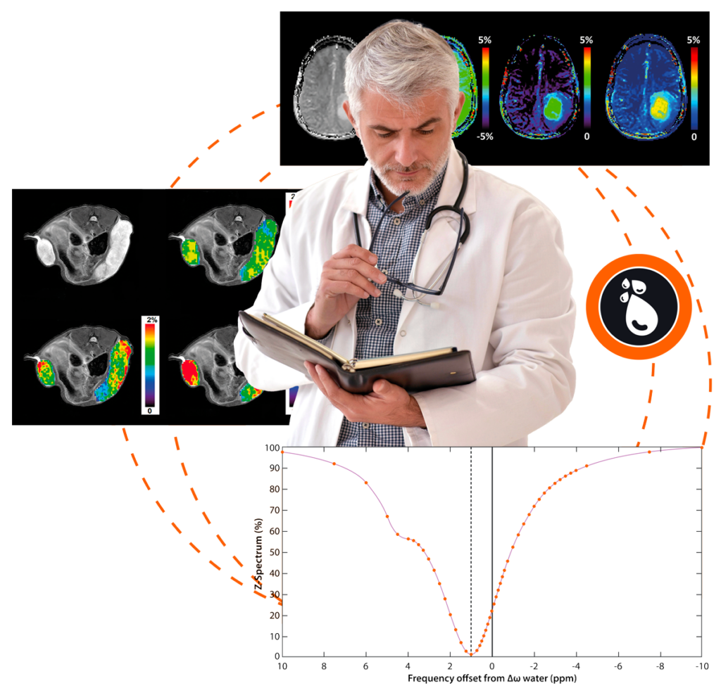
Advanced post-processing application for Chemical Exchange Saturation Transfer (CEST) MRI, specifically tailored for Amide Proton Transfer-weighted (APTw) data.

Use dedicated to research

Solution IRM APT CEST

Alignment with anatomical sequences and placement of ROIs and VOIs

Alignment of calculated maps with structural maps to accurately extract key results from VOIs and ROIs.

Different palettes and displays available for APTw maps

Adaptive map display for customized analysis, highlighting potentially anomalous areas.

Additional maps available

A comprehensive set of calculated maps providing various parameters for monitoring and analysis.

APT-CEST booklet Download
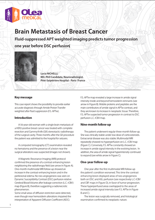
  • Olea case report: Brain Metastasis of Breast Cancer
    Brain Metastasis of Breast Cancer
    This case report shows the possibility to provide earlier accurate diagnosis through Amide Proton Transfer weighted after fluid suppression (F.S. APTw).
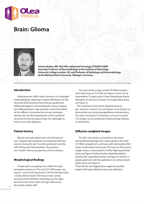
  • Olea case report: APT CEST Brain: Glioma
    APT CEST Brain: Glioma
    Multiparametric MRI in brain tumours is an invaluable methodology for obtaining in-depth information for the structural and functional tissue features guiding the differential diagnosis and assisting the tumour staging. Each MRI parametric maps provides crucial information for the different histomolecular tumour substrates, whereas the ‘tier-like’ interpretation of the combined structural-functional outputs helps the radiologist to reach an accurate diagnosis.
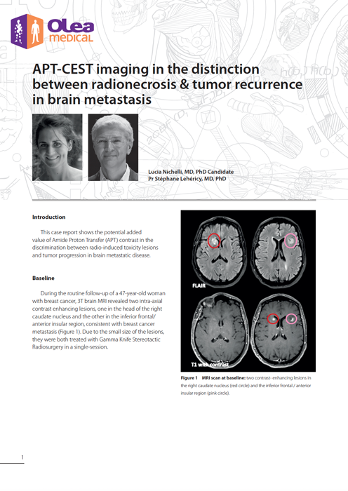
  • Olea case report: APT-CEST imaging in the distinction between radionecrosis & tumor recurrence in brain metastasis
    APT-CEST imaging in the distinction between radionecrosis & tumor recurrence in brain metastasis
    This case report shows the potential added value of Amide Proton Transfer (APT) contrast in the discrimination between radio-induced toxicity lesions and tumor progression in brain metastatic disease.
  • Amide Cs
    Amide Proton Transfer weighted imaging reveals a rolandic tumor in a pediatric patient
    The young patient presented a first hyperthermic convulsive seizure. One month later, he presented another tonic-clonic episode with left hemicorporeal seizure lasting less than 5 minutes. Another month later, he had a tonic-clonic seizure again, without fever but complicated by status epilepticus. Afterwards, a CT scan showed a right parietal lesion, and he was transferred to the Necker- Enfants Malades hospital for a brain and spinal-cord MRI. The spinal-cord MRI did not reveal any metastases.

The APT CEST solution

Present in

clinical and research centers
+ 1

Needs on average

for the post-processing of our maps
0 s

Is mentioned in

reviews and one conference article
+ 0

Overview of the main functions

APT CEST
B0 calculated map (with WASAB1, Z-Spectra or an external map)
rB1 calculated map (with WASAB1 or an external map)
B0 correction
B1 correction
Spatial noise reduction
Temporal noise reduction
Motion correction
Fluid suppression (through spillover correction)
APTw calculation at different frequency offset ranges
Alignment with the structural maps
bisdas Sotirios Bisdas, MD, PhD, MSc Neuroradiology consultant and MRI officer Department of Neuroradiology at London National Hospital of Neurology
APT-CEST imaging has become increasingly recognized as a promising imaging tool for brain tumors

Give it a go!

Request a demo or contact our sales team

The clinical and research institutes at our side

They put their trust in us

Aece9ac5 2419 4b1c 86e2 822cf55f5ccf
Hirslanden Klinik
University of Newcastle, Australia
Universitätsklinikum Heidelberg

Renseignez votre email dans le formulaire ci-dessous afin de pouvoir télécharger les case reports sur le site olea-medical