Cookie information

Cookies are small files installed on your computer or smartphone. They allow us to store information about your navigation on our site.

Privacy Policy

Cookie information

Olea Medical uses three types of cookies:

  • Session or preference cookies, which are essential for navigation and the good functioning of the site
  • Audience measurement cookies and other tracers used to establish site audience statistics

These cookies are configured according to the criteria for exemption from consent as defined by the CNIL

Cookie Type Description
Service supply Stores consent for each "tracker" type cookie.
User authentication Customer area only. This cookie will not be set if you do not log in to your account.
Service supply Stores the current language
Tracking Google analytics
Login

OLEA SPHERE® 3.0 Cardiology

One solution to assure the full continuum of cardiology MRI imaging & care
40bb8d1c8fb32ccd0e7544d4dbd68967e8d1522f

The Comprehensive, Automated and Vendor-Independent Solution

Olea Medical offers an advanced and dedicated analysis platform to get the most out of your images in a fast and efficient way.

Take advantage of the wealth of information that cardiology MRI brings

A full cardiology MRI solution for faster differenciation between normal functional areas and lesions, and analyzing cardiovascular dimensions, morphology and myocardial function.
  • Phase-contrast blood flow
  • Time signal intensity analysis
  • T2-weighted analysis
  • Integrated reporting
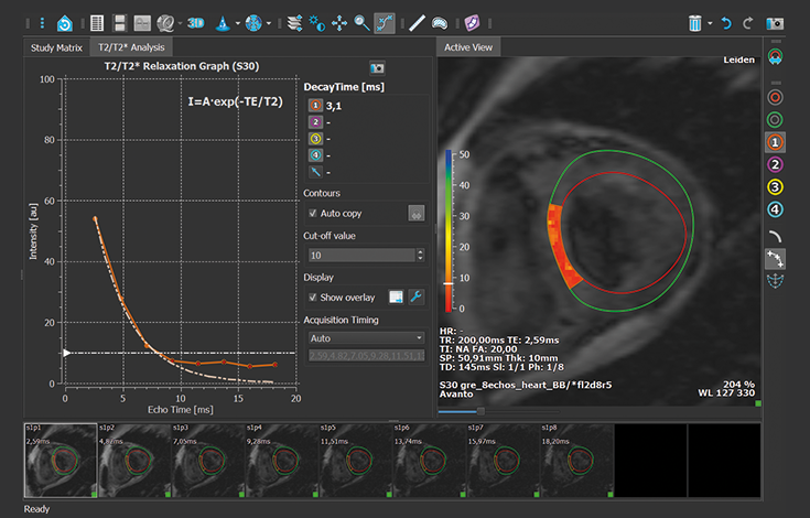
  • T2/T2* mapping, T1 mapping
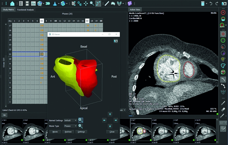
  • Volume measurement with automated contouring

One solution for visualizing the full spectrum of cardiology imaging & care

POST-PROCESS

  • Basic sculpting tools
  • T2* decay time and decay rate
  • Correct for breathing motion
  • Calculation of T1 relaxation time
  • Calculation of relative upslope and various other parameters

QUANTIFY

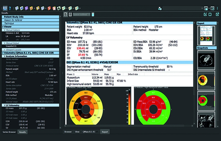
  • Left ventricle (LV) and right ventricle (RV) volumes
  • Wall motions analysis
  • Wall thickness and wall thickening measurements
  • Display of results in AHA 16 segment model
  • Display with curves of bullseye
  • Automatic infarct quantification

COLLABORATE

  • Cross-referencing tools
  • Fast paging through series
  • Export graphical report to PACS and as a PDF
  • Export numerical results via xml

ADVANCED

  • Extracellular volume
  • LV and RV Strain

"SPIN TO THE LIMIT"

Download our free book on MR Imaging

"Spin To The Limit - MRI Principles and Physical Challenges", a unique portrait of MRI,  compiled  with  the help of more  than  40  leading  experts.

Learn more

Congress

JFR 2023

Come and discover our latest solutions and meet our team at stand 206 (Hall Maillot Niv. 2)!

Learn more

OLEA MEDICAL ACADEMY

Discover our scientific webinars

More than twenty lectures, given by leading experts in the world of radiology, are available on our YouTube channel.

Learn more

Give it a try!

Request a live web demo.