Cookie information

Cookies are small files installed on your computer or smartphone. They allow us to store information about your navigation on our site.

Choose cookies Accept all cookies

Privacy Policy

Accept all cookies close

Cookie information

Olea Medical uses three types of cookies:

  • Session or preference cookies, which are essential for navigation and the good functioning of the site
  • Audience measurement cookies and other tracers used to establish site audience statistics

These cookies are configured according to the criteria for exemption from consent as defined by the CNIL

CookieTypeDescription
Service supplyStores consent for each "tracker" type cookie.
Service supplyStores authentication related to case report downloads
User authenticationCustomer area only. This cookie will not be set if you do not log in to your account.
Service supplyStores the current language
TrackingGoogle analytics

Women's imaging Mammogram and female pelvic MRI diagnosis solution
  • MRI

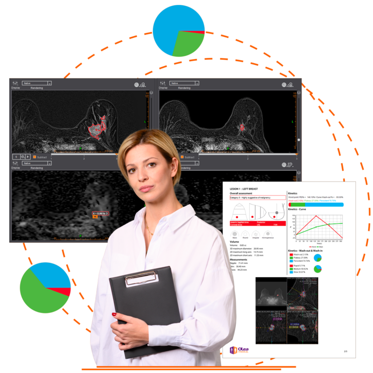
The complete solution dedicated to women’s imaging in order to make the interpretation of MRI examinations easier and improve patient management

Advanced diagnosis-assistance viewing application in breast MRIs

Developed to increase your productivity and standardize your workflow, our multi-constructor solution helps you to get the most out of your MRI examinations, while boosting your confidence in your decision-making.

Solution IRM mammaire et pelvien

Innovative applications to generalize the use of breast and female pelvic MRIs

Solution dedicated to the breast

From screening to planning biopsies, our solution integrates perfectly into your clinical practice.

Our advanced technologies allow you to quickly procure all the significant results to characterize and monitor abnormal areas of the breast.

  • Intuitive working environment dedicated to breast MRIs
  • Assistance with detecting and characterizing abnormalities
  • Segmentation of volumes and extraction of key results
  • Analysis of abnormalities enhanced by kinetic maps
  • Didactic reporting assistance (based on the BI-RADS® lexicon)
Women’s Imaging booklet Download
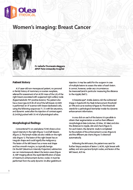
  • Olea case report: Women’s imaging: Breast Cancer
    Women’s imaging: Breast Cancer
    A 37-year-old non-menopausal patient, no personal or family history of mammary or ovarian neoplasia, referred for assessment of an ACR5 mass of the LIQ of the right breast associated with suspected right axillary node (hypermetabolic PET positive lesions). The patient also has a mass type ACR 4b of LIQ of the left breast.

Overview of the main functions

Automatic calculation of parametric maps
Analyses of morphological and functional sequences (DWI, DCE)
Automatic subtraction
4D MIP, MPR, 3D Volume rendering
Segmentation in one click
Automatic calculation of volumes and distances
Automatic measurement: lesion-to-skin, lesion-to-nipple, lesion-to-rib cage
Elastic movement correction
Longitudinal analysis
Integrated BI-RADS® report
Assistance with planning biopsies  OPTIONAL BREASTLOC MODULE (BREAST BIOPSY UNDER MRI)
Integration of grid models and multi-vendor units  OPTIONAL BREASTLOC MODULE (BREAST BIOPSY UNDER MRI)
Automatic location of the target volume of interest  OPTIONAL BREASTLOC MODULE (BREAST BIOPSY UNDER MRI)
Exportable plan report  OPTIONAL BREASTLOC MODULE (BREAST BIOPSY UNDER MRI)

3 figures to remember

breast cancer represents

new cases of cancer in women every year (3)
0 %

One woman in

will potentially develop breast cancer during her life (2)
1

By 2040

new breast cancers diagnosed, up to 3 million cases per year (1)
+ 1 %
siamotis Konstantinos Samiotis CEO & Founder at KSAM Medical Imaging Center Cyprus
breastscape solution is very ergonomic and user-friendly. All the features are very useful for me but what I really appreciate is that I am not losing my time on additional clicks, so it increases my productivity. I can examine more patients and have time for a collaborative work and that is really great!
Unknow Doc Christopher Dillon Medical Director Northwest Medical Center, Tucson, AZ
Breast MRI is an integral part of our practice CAD has always facilitated interpretations. Breastscape® runs as an application on my PACS workstation, it is simple to use and provides reliable results. There are a handful of post-processing features such as image registration, subtractions, MIP’s, and parametric maps that are most useful. Breastscape® streamlines my workflow and interpretation of cases on a daily basis.

Give it a go!

Request a demo or contact our sales team

The clinical and research institutes at our side

They put their trust in us

Clinique Olivier
Logo ERESA
Cm Albizzias
Clinic Sanador

Renseignez votre email dans le formulaire ci-dessous afin de pouvoir télécharger les case reports sur le site olea-medical