Cookie information

Cookies are small files installed on your computer or smartphone. They allow us to store information about your navigation on our site.

Choose cookies Accept all cookies

Privacy Policy

Accept all cookies close

Cookie information

Olea Medical uses three types of cookies:

  • Session or preference cookies, which are essential for navigation and the good functioning of the site
  • Audience measurement cookies and other tracers used to establish site audience statistics

These cookies are configured according to the criteria for exemption from consent as defined by the CNIL

CookieTypeDescription
Service supplyStores consent for each "tracker" type cookie.
Service supplyStores authentication related to case report downloads
User authenticationCustomer area only. This cookie will not be set if you do not log in to your account.
Service supplyStores the current language
TrackingGoogle analytics

Publications

Discover the scientific articles and publications on Olea Medical’s innovative technologies (MRI and CT), which make the difference in patient management.

Spin to the limit
Reference book on MR Imaging

“Spin To The Limit – MRI Principles and Physical Challenges”, a unique portrait of the MRI designed with the assistance of over 40 international experts.

This reference book on the MRI offers a complete overview of the past, present and future of the MRI, this unequalled imaging method that plays a central role in the coming medical revolution.

  • Predictive Imaging

    Predictive Imaging

  • Clock Around The Stroke

    Clock Around The Stroke

  • Big Bang Data

    Big Bang Data

  • Back to The Future Saga

    Back to The Future Saga

  • MRevolution

    MRevolution

  • The Diffusion Saga

    The Diffusion Saga

  • The Bayesian Saga

    The Bayesian Saga

  • Imaging Biomarkers

    Imaging Biomarkers

Predictive Imaging

Predictive imaging allows us to dream, realize the full potential of imaging, and could facilitate to achieve personalized medicine. In this 7th issue of Olea Imagein magazine, discover what experts think of the predictive value of imaging in the fields of neurology, mammography or even cardiology.

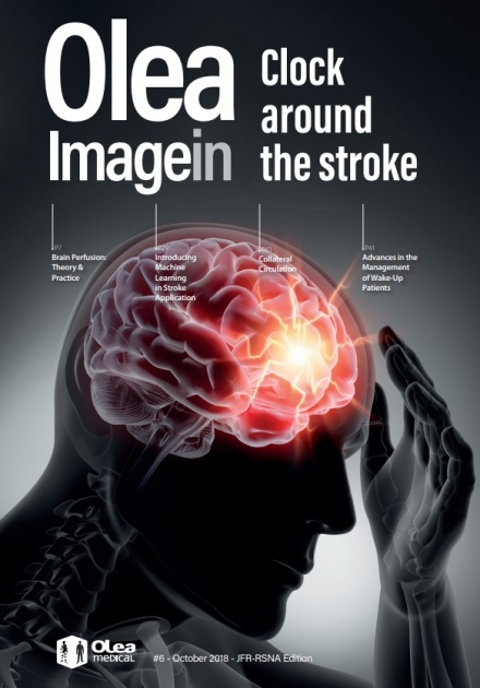
Clock Around The Stroke

Can we move from too-little-too-late to widespread timely effective care? In this issue, world leaders in the field present their opinions on key issues related to the ticking clock faced by every stroke patient. Ischemic stroke is the third leading causes of death in the world. While a slight decrease in the incidence has been observed in the past decades thanks to preventive therapies that address major risk factors such as high blood pressure, the prevalence continues to grow with the aging population.

Big Bang Data

Artificial Intelligence has grown and developed dramatically over the past few years, offering groundbreaking changes in many areas, including medical imaging. In this fifth issue of Olea Imagein magazine, broad skills experts reveal the keys to better understand and master this phenomenon. From Big Data to Machine Learning through Virtual Reality or Fingerprinting, discover the deep transformations initiated in medical imaging.

Back to The Future Saga

MRI has the highest potential to literally change the paradigm in diagnosis and patient care. Therefore, the idea behind the third issue of Olea Imagein is to relay our vision of MRI tomorrow, and the vision of experts who shape today. In this third edition, discover the Dreams of several international experts about the future of MRI, those who challenge us every day to make our and their dreams come true.

MRevolution

MRI holds a central role in the forthcoming medical revolution, the one that will transform in-depth the diagnostic and curative potential of imaging. In this 4th edition of Olea Imagein, read about the promising technologies everyone is talking about, presented by the experts who are already using them: from GlucoCEST to texture analysis, from Sindex to Kurtosis as well as from Synthetic MRI to functional MRI.

The Diffusion Saga

Diffusion imag­ing covers a broad spectrum of techniques, from the mapping of apparent diffusion coefficient (ADC) values to diffusion tensor imaging (DTI), Intravoxel incoherent motion (IVIM) or Kurtosis imaging. All these applications are based on a single physicochemical property: water motion at the cellular level. In this second edition of Olea Imagein, Olea Medical® reveals all the secrets of this technique and its potential future applications.

The Bayesian Saga

The Bayesian method is a probabilistic method based on Bayes’s theorem, published in the 18th century. Bayesian framework can be applied in any context where parameters estimation is required and noise level is a limitation. This technique represents an indisputable breakthrough and a game changer in perfusion imaging as well as diffusion and so much more. In this first edition of Olea Imagein, Olea Medical® reveals all the secrets of this unparalleled algorithm and its positive impact on both MRI and CT image post-processing.

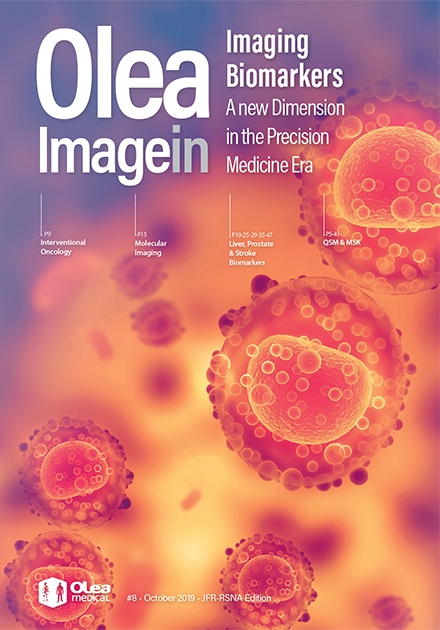
Imaging Biomarkers

Imaging biomarkers can predict patients at risk of developing a disease, detect pathologies, grade the aggressiveness of tumors and assess the response to treatment. In this 8th issue of Olea Imagein magazine, follow radiologists and experts into a new dimension, where imaging bio-markers significantly reshape the clinical research field towards precision medicine and better patient-centric care.

  • Women’s imaging: Breast Cancer

    Women’s imaging: Breast Cancer

  • Traumatic Brain Injury

    Traumatic Brain Injury

  • Prostatitis with Mild Chronic Inflammation

    Prostatitis with Mild Chronic Inflammation

  • Head & Neck: Parotid Pleomorphic Adenoma

    Head & Neck: Parotid Pleomorphic Adenoma

  • Metastatic Pancreatic Cancer

    Metastatic Pancreatic Cancer

  • Lower Limb Sarcoma

    Lower Limb Sarcoma

  • Granulomatous Prostatitis

    Granulomatous Prostatitis

  • Head & Neck: DWI and IVIM in the Orbit at 3T

    Head & Neck: DWI and IVIM in the Orbit at 3T

  • Brain: Glioma

    Brain: Glioma

  • Brain Tumor: Low Grade & High Grade Gliomas

    Brain Tumor: Low Grade & High Grade Gliomas

  • Stroke: Fronto-insular MCA occlusion

    Stroke: Fronto-insular MCA occlusion

  • Stroke: Left MCA Occlusion with Hemorrhagic Conversion

    Stroke: Left MCA Occlusion with Hemorrhagic Conversion

  • Moya-Moya

    Moya-Moya

  • MRI-Guided Thrombolysis of a Wake-Up Stroke

    MRI-Guided Thrombolysis of a Wake-Up Stroke

  • Stroke: Early Complete MCA Recanalization

    Stroke: Early Complete MCA Recanalization

  • Stroke: Left M1 Occlusion and Minor Peri-insular Infarct

    Stroke: Left M1 Occlusion and Minor Peri-insular Infarct

  • Stroke: MCA occlusion

    Stroke: MCA occlusion

  • Brain Tumor: Glioblastoma with Oligodendroglial Component

    Brain Tumor: Glioblastoma with Oligodendroglial Component

  • Brain Tumor: Low-Grade Glioma

    Brain Tumor: Low-Grade Glioma

  • Brain Tumor: Meningioma & Primary Malignant Lymphoma

    Brain Tumor: Meningioma & Primary Malignant Lymphoma

  • Brain Tumor: Pilocytic Astrocytoma

    Brain Tumor: Pilocytic Astrocytoma

  • Stroke: Right Distal M1 Occlusion

    Stroke: Right Distal M1 Occlusion

  • APT-CEST imaging in the distinction between radionecrosis & tumor recurrence in brain metastasis

    APT-CEST imaging in the distinction between radionecrosis & tumor recurrence in brain metastasis

  • APT CEST Brain: Glioma

    APT CEST Brain: Glioma

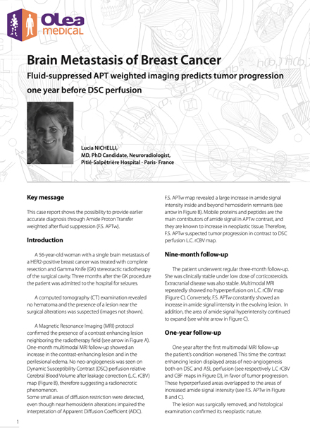
  • Brain Metastasis of Breast Cancer

    Brain Metastasis of Breast Cancer

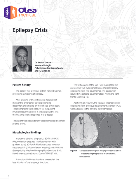
  • Epilepsy Crisis

    Epilepsy Crisis

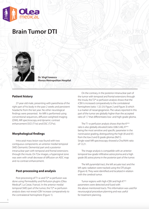
  • Brain Tumor DTI

    Brain Tumor DTI

  • Brain Cs

    Brain Metastasis of Breast Cancer

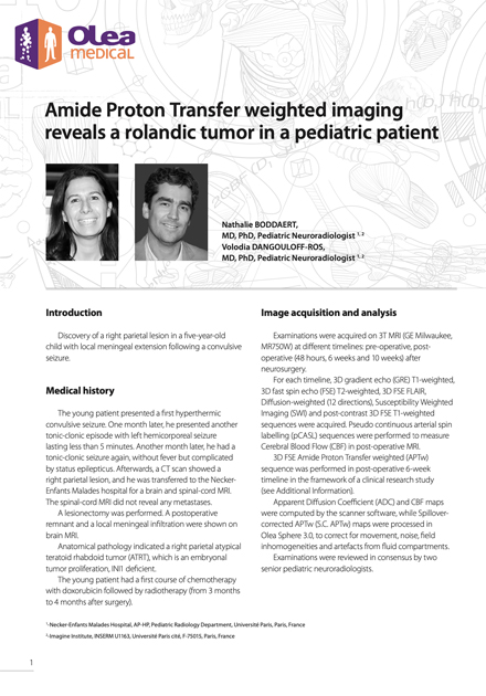
  • Amide Cs

    Amide Proton Transfer weighted imaging reveals a rolandic tumor in a pediatric patient

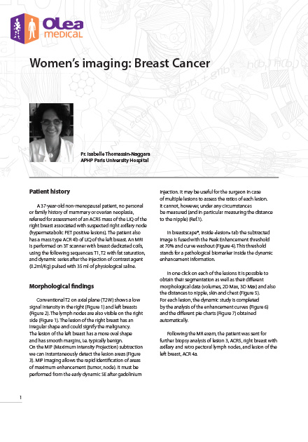
Women’s imaging: Breast Cancer

A 37-year-old non-menopausal patient, no personal or family history of mammary or ovarian neoplasia, referred for assessment of an ACR5 mass of the LIQ of the right breast associated with suspected right axillary node (hypermetabolic PET positive lesions). The patient also has a mass type ACR 4b of LIQ of the left breast.

Traumatic Brain Injury

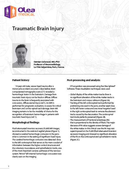
19-year-old male, severe head trauma after a motorcycle accident occurred 2 days before. Brain computerized tomography scans (CT) revealed a hemorrhagic lesion in the brainstem. Damage from traumatic brain injury can be focal or diffuse. Diffuse trauma to the brain is frequently associated with concussion, diffuse axonal injury.

Prostatitis with Mild Chronic Inflammation

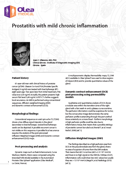
61-year-old man with clinical history of prostate cancer (pT2N0. Gleason 3+4 and PSA [Prostate Specific Antigen] 9 ng/ml) was treated with brachytherapy (BT) eight years ago. Two years later from initial treatment, PSA value was 0,39 ng/ml. Actually, the patient presents high serum PSA level: 3,49 ng/ml. A PET-CT choline suggests local recurrence. An MRI is performed using conventional sequences, diffusion weighted imaging (DWI) and dynamic contrast enhancement (DCE).

Head & Neck: Parotid Pleomorphic Adenoma

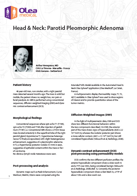
46 year-old man, non smoker, with a right parotid mass detected several months ago. The mass is solid but mobile, the patient shows no weight loss, nor pain or facial paralysis. An MRI is performed using conventional sequences, diff usion weighted imaging (DWI) and dynamic contrast enhancement (DCE).

Metastatic Pancreatic Cancer

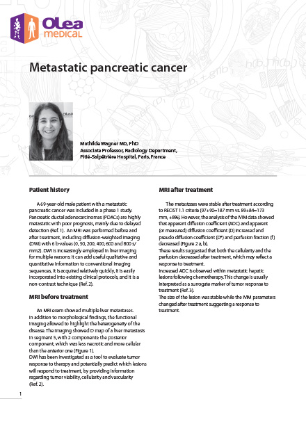
A 69-year-old male patient with a metastatic pancreatic cancer was included in a phase 1 study. Pancreatic ductal adenocarcinomas (PDACs) are highly metastatic with poor prognosis, mainly due to delayed detection (Ref. 1). An MRI was performed before and after treatment, including diffusion-weighted imaging (DWI) with 6 b-values (0, 50, 200, 400, 600 and 800 s/ mm2).

Lower Limb Sarcoma

60-year-old man, self-examination of a mass in the posterior right thigh. Referred to pre-surgery examination.

Granulomatous Prostatitis

67-year-old man with a history of recurrent superficial bladder tumor. Treatment by bladder instillations of BCG (Bacillus Calmette-Guerin) was started. Six months after the last instillation (the sixth), a rise in PSA level from 3 to 6 ng was noticed, confirmed by a repeat testing. Digital rectal examination showed a prostate firmness, predominantly on the right lobe. A prostate MRI was indicated.

Head & Neck: DWI and IVIM in the Orbit at 3T

A 38-year-old woman presented with rapidly worsening, painless monocular vision loss of her left eye, which began six months prior to the encounter. The clinical exam revealed a visual acuity of 1.4/10 and a central scotoma in the left eye. Oculomotricity was normal. An MRI was performed.

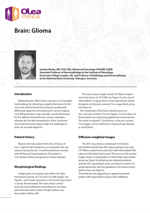
Brain: Glioma

Multiparametric MRI in brain tumours is an invaluable methodology for obtaining in-depth information for the structural and functional tissue features guiding the differential diagnosis and assisting the tumour staging.

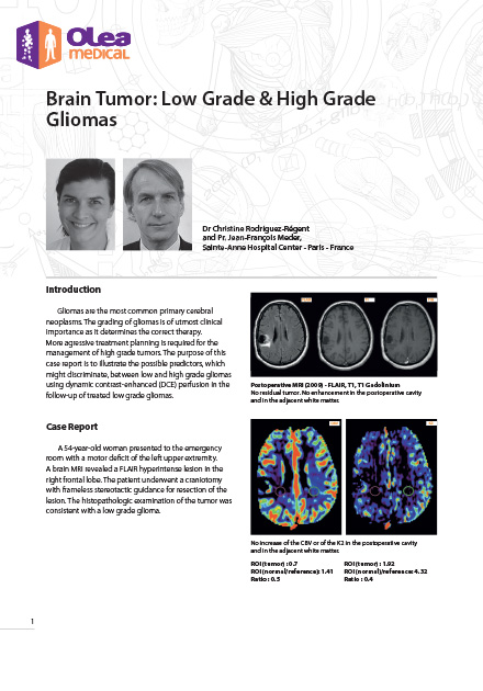
Brain Tumor: Low Grade & High Grade Gliomas

Gliomas are the most common primary cerebral neoplasms. The grading of gliomas is of utmost clinical importance as it determines the correct therapy. More aggressive treatment planning is required for the management of high-grade tumors. The purpose of this case report is to illustrate the possible predictors which might discriminate between low-and-high grade gliomas using dynamic contrast-enhanced (DCE) perfusion in the follow-up of treated low grade gliomas.

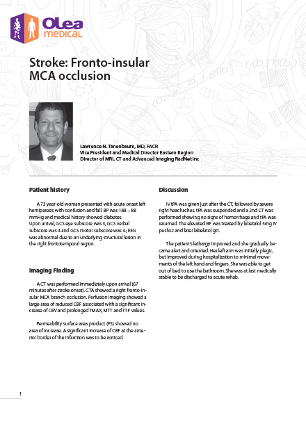
Stroke: Fronto-insular MCA occlusion

A 73yo woman presented with acute onset left hemiparesis with confusion and imbalance. BP was 188 – 88mmHg and medical history showed diabetes. Upon arrival, GCS eye subscore was 3, GCS verbal sub-score was 4 and GCS motor subscore was 4.; EEG was abnormal due to an underlying structural lesion in the right fronto-temporal region.

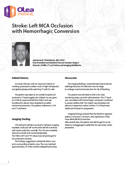
Stroke: Left MCA Occlusion with Hemorrhagic Conversion

A 64yo man with an important history of smoking presented a sudden onset of right hemiparesis and global aphasia while watching TV with his wife. The patient was taken to an the hospital and received a CT head negative for a bleed.

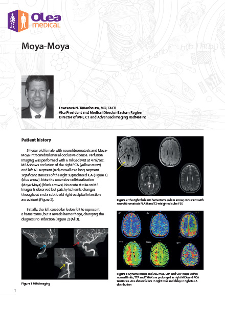
Moya-Moya

34-year old female with neurofi bromatosis and Moya Moya intracerebral arterial occlusive disease. Perfusion imaging was performed with 6 ml Gadavist at 4 ml/sec. MRA shows occlusion of the right PCA and left A1 segment as well as a long segmentsignificant stenosis of the right supraclinoid ICA.

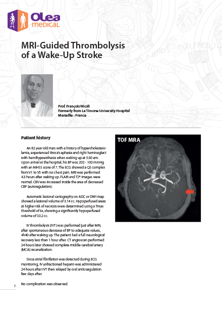
MRI-Guided Thrombolysis of a Wake-Up Stroke

An 82 year-old man with a history of hypercholesterolemia, experienced Broca’s aphasia and right hemineglect with hemihypoesthesia when waking up at 5:30 am. Upon arrival at the hospital, his BP was 200 - 100 mmHg with an NIHSS score of 7. The ECG showed a QS complex from V1 to V5 with no chest pain. MRI was performed 4.3 hours after waking up. FLAIR and T2* images were normal. CBV was increased inside the area of decreased CBF (autoregulation).

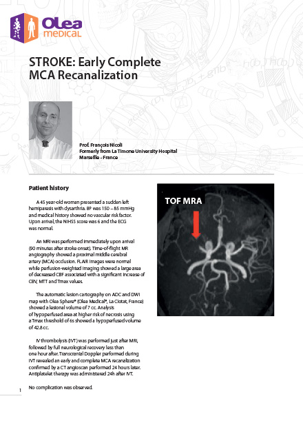
Stroke: Early Complete MCA Recanalization

A 45yo woman presented a sudden left hemiparesis with dysarthria. BP was 150 – 85 mmHg and medical history showed no vascular risk factor. Upon arrival, the NIHSS score was 6 and the ECG was normal.

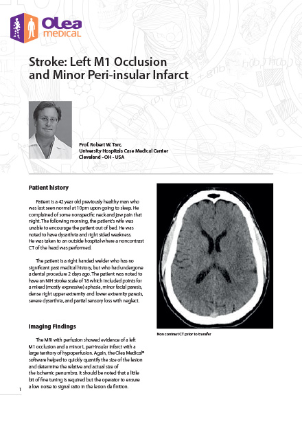
Stroke: Left M1 Occlusion and Minor Peri-insular Infarct

The patient was a 42-year-old previously healthy man who was last seen normal at 10pm upon going to sleep. He complained of some nonspecific neck and jaw pain that night. The following morning, the patient’s wife was unable to encourage the patient out of bed.

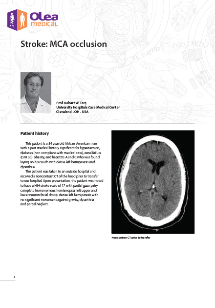
Stroke: MCA occlusion

This patient is a 54yo African American man with a significant medical history of hypertension, diabetes (non-compliant with medical care), renal failure (GFR 30), obesity, and hepatitis A and C who was found lying on his couch with dense left hemiparesis and dysarthria.

Brain Tumor: Glioblastoma with Oligodendroglial Component

A 15-year-old boy presented with headache for 1 month and vomiting in the morning for a few weeks. On neurological examination, bilateral papiloedema, bilateral paresis of the 6th cranial nerve and hemianopia were found.

Brain Tumor: Low-Grade Glioma

A 51-year-old woman with a history of LED and thyroid insufficiency, complained of continuous headache and chronic fatigue. An MRI examination in an outside hospital was performed. She then was referred to a neuro-oncologist.

Brain Tumor: Meningioma & Primary Malignant Lymphoma

This is an interesting case report demonstrating the usefulness of perfusion-weighted MRI maps for the characterization of multiple intracranial masses in a patient presenting with cerebral lymphoma.

Brain Tumor: Pilocytic Astrocytoma

This is an interesting example of how leakage correction works. The final diagnosis, pilocytic astrocytoma, is fairly straight forward. These tumors are usually described as having low cerebral blood volume, a misconception caused by the rupture of the blood brain barrier and the leakage of contrast in the interstitial medium. When corrected for agent contrast leakage the real CBV value is much higher than the uncorrected one.

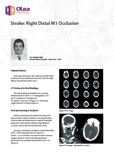
Stroke: Right Distal M1 Occlusion

An 84-year-old woman who woke up with left sided weakness. The last well-known time was 21:00 the night before. The admission NIHSS was 7.

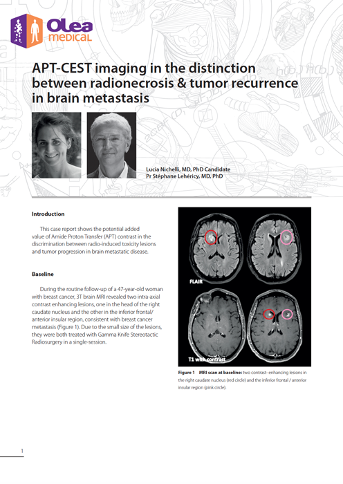
APT-CEST imaging in the distinction between radionecrosis & tumor recurrence in brain metastasis

This case report shows the potential added value of Amide Proton Transfer (APT) contrast in the discrimination between radio-induced toxicity lesions and tumor progression in brain metastatic disease.

APT CEST Brain: Glioma

Multiparametric MRI in brain tumours is an invaluable methodology for obtaining in-depth information for the structural and functional tissue features guiding the differential diagnosis and assisting the tumour staging. Each MRI parametric maps provides crucial information for the different histomolecular tumour substrates, whereas the ‘tier-like’ interpretation of the combined structural-functional outputs helps the radiologist to reach an accurate diagnosis.

Brain Metastasis of Breast Cancer

This case report shows the possibility to provide earlier accurate diagnosis through Amide Proton Transfer weighted after fluid suppression (F.S. APTw).

Epilepsy Crisis

The patient was a 40-year-old left-handed woman presenting symptoms of epilepsy. After awaking with a left brachio-facial deficit she went to emergency care experiencinq discomfort and tingling on the left side of her body. These symptoms were not new for the patient (multiple recurring events in the past) but this was the first time she had reported it to a doctor. The patient was not under any specific medical treatment prior to arrival.

Brain Tumor DTI

27-year-old male, presenting with paresthesia of the right part of his body in the past 2 weeks and persistent headache from the last year. No other neurological findings were presented. An MRI is performed using conventional sequences, diffusion weighted imaging (DWI), MR spectroscopy and dynamic contrast enhancement DCE (T1w) and DSC (T2*w).

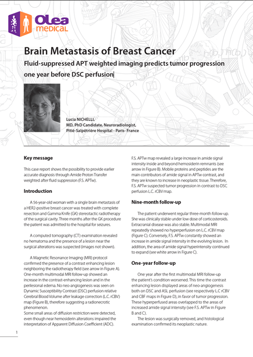
Brain Metastasis of Breast Cancer

A 56-year-old woman with a single brain metastasis of a HER2-positive breast cancer was treated with complete resection and Gamma Knife (GK) stereotactic radiotherapy of the surgical cavity. Three months after the GK procedure the patient was admitted to the hospital for seizures.

Amide Proton Transfer weighted imaging reveals a rolandic tumor in a pediatric patient

The young patient presented a first hyperthermic convulsive seizure. One month later, he presented another tonic-clonic episode with left hemicorporeal seizure lasting less than 5 minutes. Another month later, he had a tonic-clonic seizure again, without fever but complicated by status epilepticus. Afterwards, a CT scan showed a right parietal lesion, and he was transferred to the Necker- Enfants Malades hospital for a brain and spinal-cord MRI. The spinal-cord MRI did not reveal any metastases.

Subscribe to our newsletter

Add your e-mail if you would like to receive the latest news from Olea Medical.

By subscribing to our newsletter, you agree to receive news and information on Olea Medical products. Click here to view our confidentiality policy. Easy unsubscribe links are included in every e-mail.

Renseignez votre email dans le formulaire ci-dessous afin de pouvoir télécharger les case reports sur le site olea-medical